

The appearance of advertisements or/and product references in the publication is not a warranty, endorsement, or approval of the products or services advertised or of their effectiveness, quality or safety. those parts of the brain that control speech, motor functions, and senses, localization of which is important in treating brain tumors. This is particularly important when the recommended agent is a new and/or infrequently employed drug.Disclaimer: The statements, opinions and data contained in this publication are solely those of the individual authors and contributors and not of the publishers and the editor(s). However, in view of ongoing research, changes in government regulations, and the constant flow of information relating to drug therapy and drug reactions, the reader is urged to check the package insert for each drug for any changes in indications and dosage and for added warnings and precautions. No part of this publication may be translated into other languages, reproduced or utilized in any form or by any means, electronic or mechanical, including photocopying, recording, microcopying, or by any information storage and retrieval system, without permission in writing from the publisher.Drug Dosage: The authors and the publisher have exerted every effort to ensure that drug selection and dosage set forth in this text are in accord with current recommendations and practice at the time of publication. These results suggest the reproducibility, safety, and effectiveness of an early maximal functionally based resection within cortico-subcortical functional boundaries for incidental diffuse low-grade gliomas in adults in centres hyperspecialized in surgical neuro-oncology.Copyright / Drug Dosage / DisclaimerCopyright: All rights reserved.

The survival without progression requiring oncological treatment was significantly longer in patients with a total/supratotal resection than in patients with a partial/subtotal resection. Six months after surgery, all patients recovered after functional rehabilitation, with no permanent neurological deficit, Karnofsky Performance Status was 100 (except for one patient who received early postoperative radiotherapy) and no seizures were observed. Sciences (NAMS), Bir hospital were included. The resection (mean rate 96.4% range, 82.4-100) was supratotal in 5 cases, total in 5 cases, subtotal in 7 cases, and partial in 2 cases. tumors on eloquent area of brain, at National Neurosurgical Referral Center (NNRC), National Academy of Medical.
#Eloquent brain software#
We used the ANALYZE ® software (Mayo clinic, Rochester, MN, USA) to separate the brain from the overlying skull and scalp.The 120 slices of the three dimensional data set were usually edited in 90 to 120 minutes. In the immediate postsurgical period, a transient neurological worsening occurred in 10 patients. These three dimensional sequences lasted 12 minutes each. No intraoperative seizure, postoperative infection, haematoma or wound-healing problem was observed. Surgical resection of cavernous angioma located within eloquent brain areas: International survey of the practical management among 19 specialized centers. Bised a Neurosurgical Department and Departments of b Physical Medicine.

#Eloquent brain series#
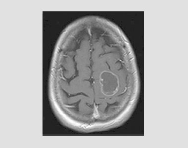
The series included 19 patients (8 men, 11 women) with no preoperative neurological deficit but with a radiologically enlarged glioma. Two-centre retrospective series of adult patients with incidental diffuse low-grade gliomas located within/close to eloquent areas in the dominant hemisphere, treated with maximal surgical resection according to functional boundaries under intraoperative functional cortico-subcortical monitoring under awake conditions, and with a minimal follow-up of 24months. We assessed the safety and efficacy of maximal safe resection according to functional boundaries for incidental diffuse low-grade gliomas in eloquent areas. Their early detection allows more effective pre-emptive and individualized oncological treatment. Incidentally discovered diffuse low-grade gliomas progress in a fashion similar to their symptomatic counterparts. The awake craniotomy is an important technique used for brain tumour excision from eloquent cortex, epilepsy surgery, and deep brain stimulation surgery.


 0 kommentar(er)
